Journal Volume 1 - January 2006
Article 8
Brain, Spinal Cord and Cells: A Neuro-primer for Non-neurologists My goal in this article is to provide you with a basic primer on the function and purpose of the brain and the spinal cord and how these organs work together. Once you have an understanding of how the spinal cord works, you can better understand transverse myelitis and how the damage to the spinal cord causes the many different symptoms of this disease. The Basic Concepts The spinal cord is part of the nervous system and facilitates the interactions between the brain and the rest of the body. The major control system is at the top (the brain) and the spinal cord acts as a bridge, communicating constantly with the brain, receiving and sending information from and to every part of the body. For example, we are able to communicate because our brain is able to generate words, and at the same time, we understand what people are saying, because our brain is processing that information. We are able to lift objects, because the brain generates the commands that go to our muscles, and these commands tell our muscles to move. All of the information going to the muscles in our limbs passes through the spinal cord. Where are the commands generated? The brain is often compared to a computer. But it is better to say that people who create computers were copying the brain, because the brain is the most amazing thing in terms of organization. The brain is a very complex structure. All of the cells called neurons are very well organized and interconnected with different areas of the brain. The brain is like a sponge. This sponge absorbs all the information from the internal environment of our body and, at the same time, collects information from our surroundings or outside environment. These brain activities are facilitated by all of the cells that are generating electrical activity. How is the brain organized? During the process of evolution, what the human brain has gained are the cells we call neurons. The increased number of these cells resulted in the gain of more and more function. That is the reason that the human brain is more complex and more capable of doing many things in terms of function, such language, speech, visual processing, memory and communication. The brain is made of gray matter and white matter. We often identify the gray matter as the cerebral cortex, but other parts of the brain also contain gray matter. Gray matter is organized in a large accumulation of neurons inside of the brain that we call gray matter nuclei (Figure 2). In the brain image obtained by MRI, we can see the cerebral cortex, the outer portion of the brain, and white matter inside of the brain. This white matter contains millions and millions of fibers that are coming from neurons and are going to establish communication with different structures of the brain. Neurons are organized mostly in structures of the brain that we call gray matter. This gray matter is shown as the outer portion of the histological section as depicted in figure 2. Neurons are organized in the brain in layers that form the cerebral cortex (gray matter). Neurons are organized in the cerebral cortex to send and receive electrical signals. These cells are organized to send information to other structures inside of the brain, which in turn send information to other parts of the central nervous system, like the spinal cord and peripheral nerves. The main center of the cell, the cell body (of the neuron), is inside the cerebral cortex or other gray matter structures of the brain that are organized in nuclei. Many of the neurons involved in the movement function of the arms or legs or any sort of motor function or modulatory function of the body organs send fibers down to the spinal cord. These fibers that are part of the neurons and facilitate communication with other neurons or organs are called axons (Figure 4). These fibers are quite important for brain function, because they facilitate the wiring system inside of the central nervous system. Axons then facilitate communication between the cerebral cortex, spinal cord and other structures in the body. A neuron is a cell; a very specialized cell that is basically comprised of a cell body in which all proteins are prepared. It is the “kitchen” of the brain. All of the chemicals and proteins necessary for the nervous system function are produced here and then transported to the end of that cell. The extension of the nerve cell is called an axon, a part of the cell similar to a fiber; the axon serves as an extension of the cell to facilitate interaction and connection with other neurons or target cells. These axons can go long distances to connect with other cells, or other neurons or other parts of the body. The length of an axon can be a few millimeters or can be longer than 50 or even more centimeters, such as the axons contained in the nerves that go from the spinal cord to our hand muscles (Figure 5). The neuron is a “chemical kitchen” This is a very fast and complex process that takes only a few milliseconds. This is the amazing feature of brain and central nervous system function. It is only a matter of milliseconds when your brain says, “move your hands;” that all of these electrical impulses, mediated by the chemical factors called neurotransmitters, take to stimulate the muscle movements. Neurons do not work alone. There are other cells in the nervous system, glial cells and blood vessels that coexist with neurons that maintain the function of the brain. There are specific glial cells that are particularly important in brain function. One group of glial cells, the astrocytes, are available to support neuronal function and also facilitate communication with the blood stream. Remember, the blood stream is very important for brain function and spinal cord function, because all nutrients are coming to the brain through the blood stream. The blood stream facilitates nutrients to neurons, a function that is facilitated by astrocytes through regulation of the blood-brain barrier. Another type of glial cell is the oligodendrocyte (Figure 4). The oligodendrocyte is the factory for myelin production within the brain and spinal cord. Myelin is an important part of the axon. Myelin is a wrapping around the axon like the insulating wrapping around an electrical cord (Figure 4). In the brain and in the nervous system, in general, the myelin covers a major portion of the axons. Oligodendrocytes are the factories which constantly produce this myelin, the “wrapping” around the axon, and are extremely important for the survival of the neuron. What about the spinal cord? The spinal cord is the main highway that facilitates communication between the periphery, the arms, legs, nerves, skin and the brain. Many receptors for pain and many receptors for different types of stimulations that are on the skin are communicated with the spinal cord through this very fine structure called peripheral nerves. We have peripheral nerves everywhere and they also act as bridges between the skin or the periphery and the spinal cord. There is a constant communication with the spinal cord and eventually with the brain by using all of these small fibers that are in the periphery. At the same time, the brain is sending a lot of information downstream. This information is coming downward from the cerebral cortex to the spinal cord and getting organized in the spinal cord to facilitate different functions. Some of these functions being organized in the spinal cord are motor activity; information that will eventually go to different muscle groups in the arms or legs (Figure 7). At the same time, information from the periphery arrives to the spinal cord, where it is organized and transmitted to the brain as part of sensory information. As is the case with the brain, the spinal cord is extremely well-organized. The spinal cord is also organized into gray matter and white matter compartments, but is organized differently than in the brain. In the brain, the gray matter is the outer portion of the brain. In the spinal cord, the gray matter is located in the inner part of the cord (Figure 8). Gray matter is basically an accumulation of neurons that deal with either motor or sensory function. The neurons localized in the anterior portion (ventral horn) of the gray matter in the spinal cord are in charge of motor function while the neurons located in the posterior area of the cord gray matter are dealing with sensation. The neurons in the ventral horn receive commands from the motor cortex in the brain and they generate the commands that are going to tell the arm or leg muscles to move. These commands are facilitated by the information that is traveling through “motor” peripheral nerves, a package of axons from motor neurons. These axons will connect motor neurons with the end targets, the muscles. The neurons of the posterior region (dorsal) of the gray matter in the spinal cord are involved in sensory function. These are the neurons that receive information from the periphery through “sensory” peripheral nerves. Again, these nerves are packages of axons, but in this case coming from peripheral receptors on the skin, muscles or other organs. The outer portion of the spinal cord is the white matter. If you remember, white matter contains the wiring for communication between the brain and other structures and vice versa. In the spinal cord, the white matter is on the outer compartment and it is organized in downward pathways coming from the brain down (descending tracts), or in upward pathways going from the spinal cord to the brain (ascending tracts). This white matter in the spinal cord is also well organized, so ascending and descending pathways are distributed in specific areas of the cord (Figure 9). For example, in the lateral portion of the spinal cord, we have a highway of motor function coming from the brain to the spinal cord. This highway is called the “cortical spinal tract” and the name cortical is derived from the fact that this tract is connecting the cortex with the spinal cord. These are the major tracts that carry information for motor function. When there is involvement of the lateral portion of the spinal cord, that damage will produce disruption of communication between brain and spinal cord resulting in lack of motor commands for movement and motor function. There is another type of information that is going upward to the brain. Those are basically pathways that we call ascending highways. These ascending highways carry information that has been collected from other parts of the body, organs or skin to the brain or other parts of the central nervous system. What happens when there is injury to the brain or spinal cord? There are many areas of the neuron, and many parts of the neuron that are susceptible to different types of attack. Myelin is particularly important, because in the case of multiple sclerosis, the immune reactions that occur in that disease are going to target specifically the myelin. There are other disorders in which the main target of attack is the cell body. For example, in Lou Gehrig’s disease, the main problem is the cell body of motor neurons in the spinal cord. For unknown reasons, this disease can start a process of degeneration of that cell body. In other diseases, like in peripheral neuropathies, the main problem is either the myelin or the axon. So again, the neuron has a number of different parts and these different parts can be affected by different types of disorders. In some diseases of the brain and spinal cord, the only part of the neuron that is affected is the myelin. In the case of multiple sclerosis or other types of immunological attacks against the nervous system or the spinal cord, myelin is one of the main targets of “inflammatory” attacks. But in some diseases, the attack may be more extensive; the myelin is attacked, the axon is attacked, and the cell body is attacked, so the consequences are grave as the possibility of cell death increases. There is another problem that I will mention briefly. In addition to the type of immune attack that I have just described, there is a possibility of strokes in the spinal cord. Lack of blood supply to the spinal cord will generate a lot of distress to the neurons. Due to the lack of oxygen, neurons will stop the production of proteins that maintain neuronal survival. So strokes or lack of blood supply to the spinal cord may produce irreversible damage to the neurons. What is the meaning of spinal cord anatomy and TM? Damage to the ascending pathways translates into disruption of the information going to the brain: SENSORY FUNCTION Symptoms:
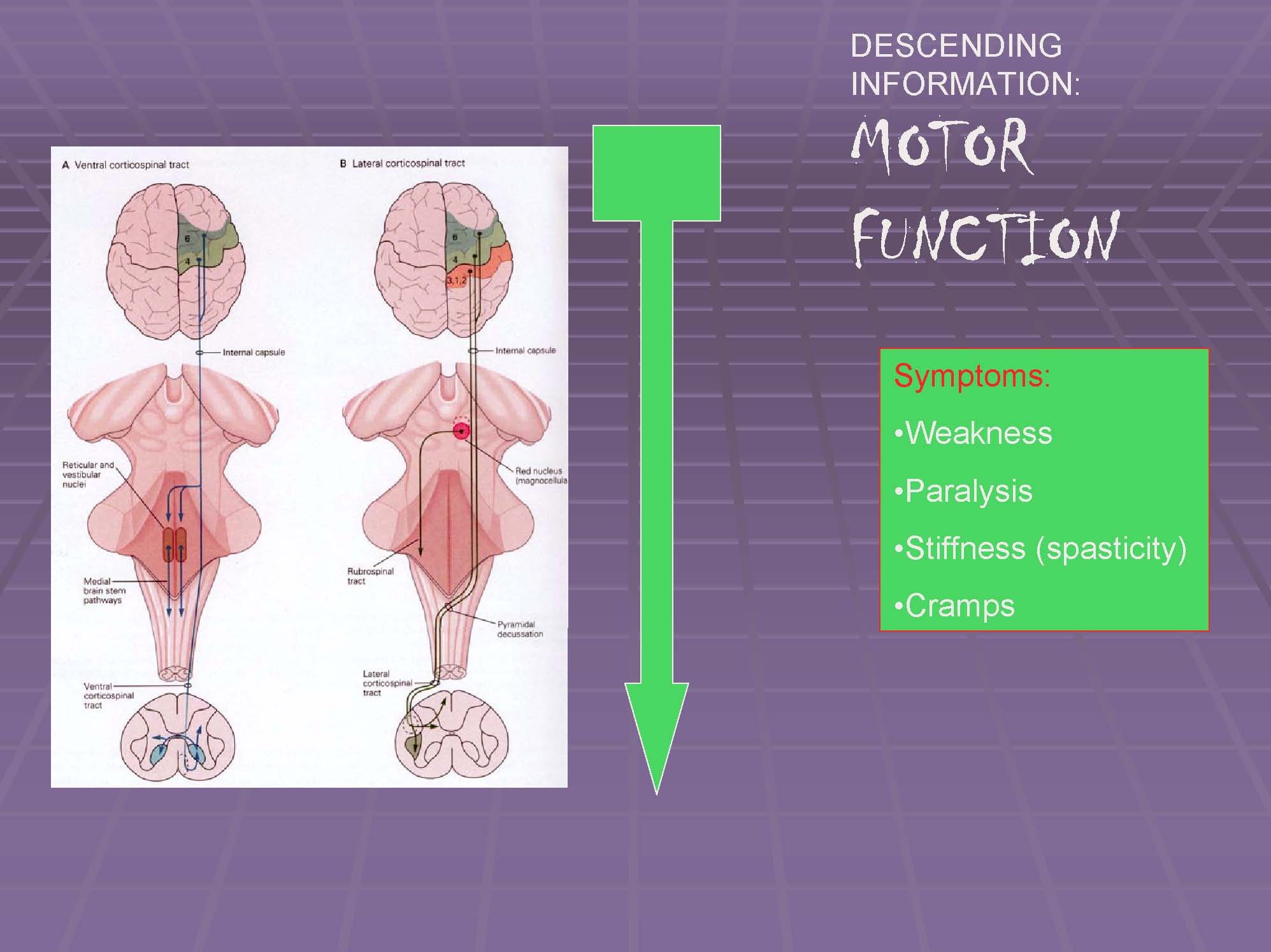
For example, every time you touch something, your nervous system connects a network that consists of receptors that you have in your finger that are going to the dorsal root ganglion where there are neurons that establish communication with the spinal cord. This sensory communication comes to the posterior gray matter in the spinal cord. When you have this communication, the spinal cord is going to tell the brain, “my finger is detecting some signals,” and that signal is going upward to the brain and establishing connections with other portions of the brain and with portions of the cerebral cortex (Figure 10). So, this is a very well organized communication. This is a network that works in milliseconds, and it is working by using many chemicals and many electrical activities. When we disturb this sensory function (through an immune attack or through a spinal stroke or through a traumatic injury), we may experience pain, lack of sensation, abnormal sensation or lack of balance. This is an important concept, because many patients with transverse myelitis (TM) complain about the lack of balance. Even in patients without muscle weakness, there is a possibility of balance and sensory problems because of the potential involvement of those specific areas of the spinal cord dealing with sensory information. This situation is caused by the central nervous system being unable to receive communication from the periphery; this communication is disrupted by the damage caused to the spinal cord that occurs in TM. Another sensory symptom in transverse myelitis is pain. This is one of the most excruciating and frustrating situations for our patients. Pain originates either in the periphery or in the spinal cord. In patients with transverse myelitis, we believe that the main center of pain activity is in the spinal cord, and not in the periphery. Many patients complain about burning sensations in their feet and other uncomfortable sensations. These pain and sensory problems may occur in different parts of the body and the distribution of pain depends on the part of the spinal cord that is affected. The symptoms originate when the spinal cord that is undergoing inflammation, damage or abnormal electrical activity, generates abnormal and dysfunctional “firing” of the neurons connected with sensory and pain information. Many times, even when the inflammatory activity or injury has subsided, pain remains because there is either permanent damage to the networks associated with pain or because there is abnormal re-wiring of the networks. This latter factor originates from the attempts for regeneration of the spinal cord that follow the injury. Often, the process of regeneration induces abnormal electrical-chemical activity that translates into pain. In many ways, pain in patients with transverse myelitis is a bad thing to have. But in other ways, it is a reminder that something is going on in the spinal cord, and perhaps there is some type of regenerative activity in the spinal cord that is inducing that pain. Damage of the descending pathways translate into disruption of the information going from the brain to the spinal cord: MOTOR FUNCTION Symptoms: Now, let’s turn to a discussion about motor function. Motor function is initiated from signals that come from the brain and travel to the spinal cord. That function is well localized in a portion of the cerebral cortex that is called the motor cortex. This information travels downwards through two different highways. The main highway, the corticospinal tract, is a big package of axons that carries a lot of “traffic” information between one part of the brain (the brain hemisphere) and the spinal cord on the contralateral side (Figure 11). In this way, the right brain is moving the left side of the body and the left brain is moving the right side of the body. That happens because this highway crosses to the other side in the portion of the brain that is called the brain stem. All of this motor function information is carried from the motor cortex in the brain to the spinal cord, and then from the spinal cord to the muscles; a complex communication that translates into the production of movement. When this communication is disrupted because of transverse myelitis, the immediate consequence is weakness, or the most extreme of weakness, which is paralysis. The disruption of motor function that is caused by TM may also result in lack of muscle tone (hypotonia) or stiffness, symptoms that are frequently associated with weakness. The stiffness is what we call in medical terms, spasticity. The variability of symptoms in TM depends on the location of the lesions Damage of the white matter: Motor and sensory symptoms The medical term for problems with balance or the inability to coordinate muscle movements is ataxia. These type of symptoms may present alone or in combination with other sensory abnormalities or movement ( motor) dysfunction that may be associated with damage of the lateral white matter pathways (descending, motor information) (Figure 12b). An example of a complex and aggressive situation is when TM affects both gray matter and white matter compartments simultaneously (Figure 13a and Figure 13b). These are the catastrophic situations that we have in some patients with transverse myelitis. There is no motor function; there is complete paralysis, spasticity and absence of the different sensation modalities. These are, unfortunately, extreme cases of attacks against the spinal cord, because in many cases, the destruction of the gray matter is often an irreversible process and the neurons that are dying are not going to be regenerated by the spinal cord. Multiple sclerosis, for example, produces an attack against the white matter, but in many patients with transverse myelitis the attack may extend beyond the white matter. Damage in this central portion of the spinal cord will cause a combination of motor dysfunction with sensory dysfunction. Conclusion |